Join Our Team! View Open Positions
CT
For compassionate, high-tech imaging services, make Orthopedic Associates of Lancaster your trusted source. Extremity Cat Scan (CT) appointments are now available at our Lancaster office. Call 717-299-4871 to schedule an appointment.
Thanks to CT scanning (computed tomography, often called a “CAT” scan) doctors can view a “slice” of the human body painlessly. Orthopedic Associates of Lancaster offers extremity CT scans for the upper and lower extremities, allowing faster scan times and quicker results back to your doctor for follow up.
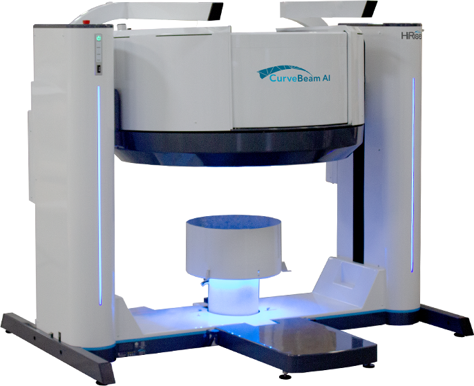
At Orthopedic Associates of Lancaster, we offer patients advanced state-of-the-art imaging technology through the CurveBeam AI HiRise Extremity CT Scanner.
HiRise Extremity CT
The state-of-the-art HiRise extremity CT scanner at Orthopedic Associates of Lancaster also offers a Metal Artefact Reduction (MAR) feature for patients that may have already had surgical intervention, which allows for quality imaging even when metal is present in the desired joint.
The HiRise extremity CT scanner uses a very low dose of radiation compared to a normal CT scanner, allowing less overall patient dose and still very high-quality images. This scanner allows doctors to assess new and healing fractures as well as pre-operative planning for upcoming surgeries. The scanning process takes approximately 10 to 15 minutes in total.
CurveBeam AI CT
CurveBeam AI is a technology utilizing standing CT scans for detailed 3D imaging of your bone alignment and joint spaces. This is done by acquiring and combining multiple X-ray projections to create a 3D representation of the patient’s body.
In addition to being a comfortable option for necessary diagnostics, a scan in the standing position allows our doctors to easily evaluate the alignment of bones and joints. It also gives the most accurate assessment of the function of the affected area for weight-bearing joints.
Standing CT scans can be as a diagnostic tool for:
- Pre-operative planning
- Post-operative assessment
- Diagnosis of fractures
- Evaluation of arthritic joints
- Evaluation of bunion deformity in the foot
- Evaluation of patellar instability in the knee
- Evaluation of leg instability
- Evaluation of leg alignment
- Bone position and condition
LOWER EXTREMITY
Lower extremity scans include feet, ankles, knees, and standing hips. Allowing the patient to remain standing for the test results in weight-bearing images for the physician to review. These images differ from standard CT scanners and help inform the physician of possible alignment-determinant conditions and create a better assessment of a patient’s gait under normal standing pressure. Some popular indications for a weight-bearing CT exam include Lisfranc, Hindfoot alignment evaluation, and Charcot. We are also able to perform these studies non-weight-bearing (excluding hips) due to patient condition or recent injury if necessary.
Please note: We do not perform scans on knees with full replacements regardless of whether it is the affected side or unaffected side.
- Foot (weight-bearing or non-weight-bearing)
- Ankle (weight-bearing or non-weight-bearing)
- Knee (weight-bearing or non-weight-bearing)
- Hip (weight-bearing)
UPPER EXTREMITY
Upper extremity scans include hands, wrists, and elbows. The flexibility of this machine allows patients to sit during their exam, creating a more comfortable atmosphere when trying to capture the best image possible. The patient’s extremity is inserted straight into the machine, so there isn’t any awkward positioning or pain for the patient.
- Hand
- Wrist
- Elbow